what nerve carries the impulses to the brain
The brain and spinal string make up the central nervous system. The spinal cord, simply put, is an extension of the brain. Information technology is an ovoid shaped column of nervus tissue that extends from the brain down to the 2nd lumbar vertebrae. It allows us to command our arms, legs, and our bathroom habits, among many other things.
The spinal cord is enclosed in protective tissues chosen the meninges. The meninges form a protective sack effectually the spinal cord. Inside the spinal (ordural) sac, the spinal cord is surrounded by a nourishing fluid called cerebrospinal fluid. The dural sac is further protected past the bones of the spinal column.
The internal anatomy of the spinal cord is quite complex. To keep things unproblematic, the center of the cord consists of gray mater. White mater is arranged in tracts around thegray mater. It consists of axons that transmit impulses to and from the brain or betwixt levels of grey mater within the spinal cord.
The spinal cord has two basic functions. The spinal string carries sensory impulses to the encephalon (i.due east. allows united states of america to feel) and motor impulses (i.eastward. allows u.s.a. to move our muscles) from the brain. The spinal cord also controls stretch reflexes and controls our bowel and bladder functions.
The spinal cord also acts every bit a nerve centre betwixt the brain and the rest of our trunk. Thirty-i pairs of fretfulness exit from the spinal string to innervate our body.
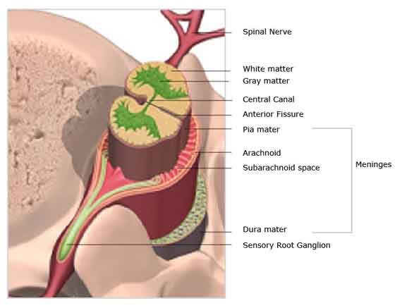
Labeled Cross Section of Spinal Cord

- Anterior Fissure
- Deep groove along the forepart of the spinal string
- Meninges
- The three membranes that cover and protect the spinal string and encephalon—dura mater (outer), arachnoid (middle) and pia mater (inner)
- Pia Mater
- This sparse vascular membrane of collagen fibers is the innermost layer of the iii meninges that covers and protects the spinal cord and encephalon.
- Arachnoid
- The middle layer of the 3 meninges that covers and protects the spinal cord and brain.
- Subarachnoid Infinite
- The surface area betwixt the arachnoid membrane and the pia mater
- Dura
- The outermost, toughest, and nigh fibrous of the three membranes that covers and protect the spinal cord and brain
- Greyness Mater
- Gray mater is divided into three functional zones— the dorsal horns (sensory), the ventral horns (motor), and the eye zone (links the two horns). Grey mater is made up of neurons which are motor or sensory in nature.
- White Mater
- White mater consists of axons of neurons grouped in bundles that incorporate nervus fibers. These bundles travel between the spinal cord and the brain. Pathways to the brain are usually sensory, and pathways from the encephalon to the spinal cord are normally motor in nature.
- Sensory Root
- Collection of the cell bodies of the sensory nerves.
- Spinal Nervus
- Sensory and motor nerve rootlets merge to grade a spinal nerve
Spinal Fretfulness
In that location are 31 pairs of spinal nerves that arise from the spinal cord. Each spinal nerve corresponds to the level it emerges from: there are 8 cervical, 12 thoracic (chest), five lumbar (lower back), and 5 sacral, and 1 coccygeal (tailbone) nerves. Each spinal nerve is fastened to the spinal cord past 2 roots: a dorsal (or posterior) sensory root and a ventral (or inductive) motor root.
The fibers of the sensory root bear sensory impulses to the spinal cord —pain, temperature, touch and position sense (proprioception)—from tendons, joints and body surfaces. The motor roots carry impulses from the spinal cord to the muscles. The spinal nerves exit the spinal cord and pass through the intervertebral foramen.
Nerve Plexus
A nerve plexus is a network of multiple nerves. The spinal fretfulness in each part of the spine cluster together to form a plexus. Listed below are the named plexuses:
- Cervical plexus
- A network formed by the first iv cervical spinal nerves. It innervates parts of the face up, neck, shoulder and chest and gives rise to the phrenic nervus which controls the diaphragm (allows u.s.a. to jiff).
- Brachial plexus
- A network of the final iv cervical and first thoracic spinal fretfulness. These nerves supply the shoulder, arm, forearm and mitt.
- Lumbosacral plexus
- A network of lumbar and sacral nerves which supplies the lower extremity.
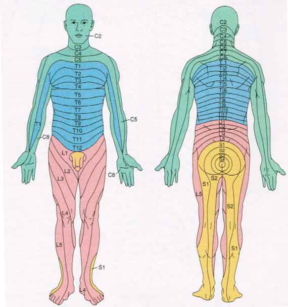
Map of Dermatomes
A dermatome is a band or region of pare supplied by a unmarried sensory nerve. Sensory nerves carry sensory impulses to the spinal string. Sensory impulses include pain, temperature, touch and position sense (proprioception)—from tendons, joints and body surfaces. Every office of the body has a dermatome that is supplied by a spinal nerve. The exception to this rule is the face, which is supplied past the cranial fretfulness.

Key to Spinal Nerve Regions
Each pair of spinal nerves links to one of 4 regions of the body.
- Cervical Region (greenish): 8 pairs of nerves supply the skin covering the back of the head, the cervix, shoulders, arms and hands.
- Thoracic Region (blue): 12 pairs of thoracic nerves supply the skin on the breast, dorsum and nether artillery
- Lumbar Region (pinkish): 5 pairs of lumbar nerves supply the skin on the lower abdomen, thighs and fronts of the legs
- Sacral Region (yellowish): Sacral nerves supply the pare on the rears of the legs, the feet and genital areas
Levels of principal dermatomes
- C5 — clavicles
- C5, C6, C7 — lateral parts of the upper limb
- C8, T1 — medial sides of the upper limb
- C6 — pollex
- C6, C7, C8 — mitt
- C8 — ring and little fingers
- T4 — level of nipples
- T10 — level of umbilicus
- T12 — inguinal or groin regions
- L1 L2 L3 L4 — anterior and inner services of lower limb
- L4, L5, S1 — foot
- L4 — medial side of large toe
- S1. S2, L5 — posterior and outer surfaces of lower limbs
- S1 — lateral margin of human foot and little toe
- S2, S3, S4 — perineum
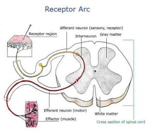
Receptor Arc

In the nervous system there is a "closed loop" system of sensation (sensory), decision (brain), and reactions (motor). This process is carried out through the activeness of afferent neurons (sensory), interneurons (spinal string), and efferent (motor) neurons. This closed loop forms a receptor reflex arc. While near nervus stimuli are processed through the brain, this is receptor reflex arc is processed at the level of the spinal string to allow for a lightning fast response.
Receptor reflex arcs allow for automatic responses to dangerous stimuli (hurting, abnormal positioning of a limb) before you would otherwise think to respond to them. An example of this is the automatic withdrawal response that occurs when yous accidentally touch something that is very hot. The receptor arc automatically responds by withdrawing your arm, earlier y'all can fifty-fifty call back to do and so — i.e. before you can even procedure that you've touched something hot.
More information well-nigh your nerves, spine and dorsum
- Anatomy Terms
- Lumbar Spine Beefcake (lower back)
- Spinal Cord Beefcake
- Spinal String Injury
Source: https://www.healthpages.org/anatomy-function/spinal-cord-anatomy/
0 Response to "what nerve carries the impulses to the brain"
Post a Comment